Abstract Clinically, the treatment of patellar dislocation in dogs is mainly treated through trolley wedge or rectangular resection, quadriceps femoris release, joint capsule release, support ligament relaxation, tibial tuberculosis displacement, jo...
Abstract
Clinically, the treatment of patellar dislocation in dogs is mainly treated through trolley wedge or rectangular resection, quadriceps femoris release, joint capsule release, support ligament relaxation, tibial tuberculosis displacement, joint capsule overlap, corrected osteotomy, and trolley replacement. However, due to the different incidence and complexity of different dogs, various treatment methods have certain limitations. This article reports a case of Teddy dog with bilateral hindlimb lameness. It was diagnosed with bilateral lateral patellar dislocation by X-ray. It was treated with tracheal groove rectangular resection, tibial tubercle displacement and articular capsule overlapping surgery, and was basically cured after 1 month.
Background
The patella is also called the kneecap, which is a small bone under the extensor of the quadriceps femoris. Clinically, patellar dislocation is often found in small dogs, and sometimes in large dogs. Patellar dislocation has a 7.2% incidence in all genetic diseases, and a higher incidence in purebred dogs. The causes of most patellar dislocations are abnormal skeletal muscles, such as intramotor movement of the quadriceps femoris, rotation of the distal femoral lateral, curvature of the 1/3 of the distal femoral, dysplasia of the femoral shaft, unstable knee rotation and deformation of the tibia. Patellar dislocation is generally divided into four levels in clinical practice: Level I - intermittent patellar dislocation, and spontaneous dislocation rarely occurs during normal exercise. Artificial patellar dislocation occurs when the knee joint is bent during the physical examination, but when the pressure is discharged, the patella is reset and the joint flexion and stretch return to normal. Grade II - The femur is torsion and slightly deformed. The patella is dislocation under the action of artificial external forces such as squeezing or flexing the knee joint. It can only be reset when the opposite force is applied and the tibia of the sick dog is reversely rotated. Grade III - The patella is in a dislocation state in most cases and can only be manually reduced when the knee joint is stretched. However, when resetting successfully, it will be dislocated again as the knee joint flexes, stretches and moves. Grade IV - The proximal tibial platform rotates from 80° to 90°, and the patella cannot be reset manually. The trochanter groove at the distal femur becomes shallow or disappears, the quadriceps femoris displaces, the soft tissue structure supporting the knee joint is abnormal, the femur and tibia are deformed significantly, and clinically manifests symptoms such as lameness, difficulty in carrying weights, and curvature of the affected limbs.
Case data
1 male teddy dog, 2 years old, weighing 6.2kg, did not undergo castration surgery, fully immunized, and kept indoors. The affected dog's bilateral hind limbs are limping for half a year, and it will have a "rabbit jump" posture when running.
Clinical examination
Body temperature was 39℃, pulse was 115 times/min, diet and stool were normal. Place the sick dog on a soft blanket with a belly facing up, hold the distal femur of the left affected limb with your left hand, and hold the distal tibia with your right hand, and find that the affected limb cannot be stretched by knee flexing. The thumb and index finger of the left hand pressed against the knee joint, flexed the knee joint, palpated the patella and found that the patella was not inside the troika. After careful examination, a free lump was found on the outside of the troika. Artificial force pushing the hard block inward can be reset, and the leg will be released again when it bends its knees. The palpation trough is smooth, there is a "knock" sound when bent your knees, and there is a significant pain. The same method was used to examine the right affected limb, and the results were the same, and the lateral dislocation of the patella was highly suspected.
Imaging examination
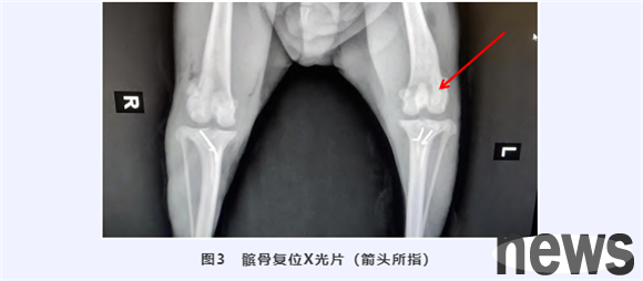
After X-ray examination, the bilateral trochantergic groove of the affected dog became shallow and the bilateral lateral patella was tertiary dislocation (Figure 1).
Surgery
1) The surgical pathway is opened. Anterior lateral skin incision was made from 4 cm proximal to the patella and extended 2 cm below the tibial tubercle. Incite the subcutaneous tissue along the same line, incite the lateral support ligament and joint capsule to expose the joint.
2) Tackle rectangular resection. Cut the articular cartilage of the trochlear to have a rectangular appearance to ensure that the width of the incision is sufficient to accommodate the width of the patella, but keep the trumpet crest. Small and medium-sized dog cases use a hand-held saw to cut the bone 2~6mm, and use an osteoch of the same width as osteotomy to lift the bone cartilage block (Figure 2). The osteochi was inserted from the proximal and distal ends of the osteotomy and converged in the middle. Carefully remove bones of appropriate thickness and avoid cracking of bone cartilage blocks. Remove excess bone from the base of the groove to deepen the trench. When the depth is sufficient to accommodate 50% of the patella height, the free osteocartilage block is reset. Rinse away the blood clots and epiphysis with normal saline, and then re-cover the bone cartilage block in the trench groove to form a new cartilage joint. Due to the compression force of the patella and the friction of the cancellous bone surface on the cross-section, the bone cartilage block can be kept in place.
3) Tibal tuberculosis dislocation. By incision through fascia lata on the lateral side parallel to the patella, the incision extends distally to the tibial tuberculosis below the joint line. The anterior tibial muscle is lifted from the lateral side of the tibial tuberculosis and from the tibial platform. Sharp separation, obtaining a deep-level pathway of the patellar tendon to facilitate the removal of the osteotomy. The width of the nodule resection should be sufficient, the caudal to the muscular groove should be kept intact at the distal osteotomy. At this time, the surgeon should choose the appropriate attachment point for the tibial tuberculosis, and usually use 2 K-Spots to refix the tibial tuberculosis. The first K-Shitcher needle is perpendicular to the osteotomy plane, and the second K-Shitcher needle is perpendicular to the straight knee ligament. After the tibial tubercle displacement surgery is completed, the blood clots in the surgical area are rinsed with a large amount of sterilized saline, and the knee joint is flexed to check its stability.
4) Close the surgical wound. The excess joint capsule is removed, and the fascia of the joint capsule and knee joint is nodularly sutured using absorbable sutures in the first layer, and then the proximal fascia of the patella is continuously sutured, the muscle layer is continuously sutured layer by layer, and the skin is nodular sutured. Rinse the wound with saline or chlorhexidine, clean the blood stains near the wound, and finally wipe the wound with iodine.
Discussion
Because the patella is in a dislocation state for a long time, the support ligament on the opposite side of the dislocation will be stretched, and the excess support ligament and joint capsule will be removed, which will make the closed joint incision tighter. When the rectangle of the trench groove is deepened, cutting too close to the edge will cause fracture of the trench ridge; when the trench trochanter is displaced, the cutting point should be in front of the muscular groove. If the trench trochanter is accidentally cut completely, the trench tubercle should be fixed with the "8" wire.
There are many physiological and pathological factors for patellar dislocation in dogs, and suitable surgical plans should be selected for different cases.. Normally, grade I patellar dislocation does not require surgical treatment. Most affected dogs can be well controlled through conservative treatment and long-term oral treatment of Purvecon; grade II to III patellar dislocation requires the use of trolley deepening and tibial trochanteral dislocation to correct. Simple trolley deepening surgery will cause a quick recurrence after the operation and cannot achieve good treatment effect; grade IV patellar dislocation requires the choice of trolley deepening, tibial trochanteral dislocation and osteotomy or trolley ridge thickening according to the situation. In addition, arthritis caused by patellar dislocation itself and pain caused by surgery to damage to joint cartilage requires long-term oral shark chondroitin or regular oral nonsteroidal anti-inflammatory drugs for pain management.